Mi On Ecg

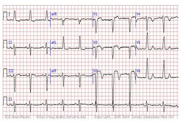
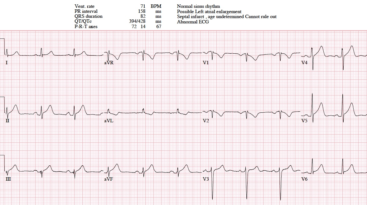
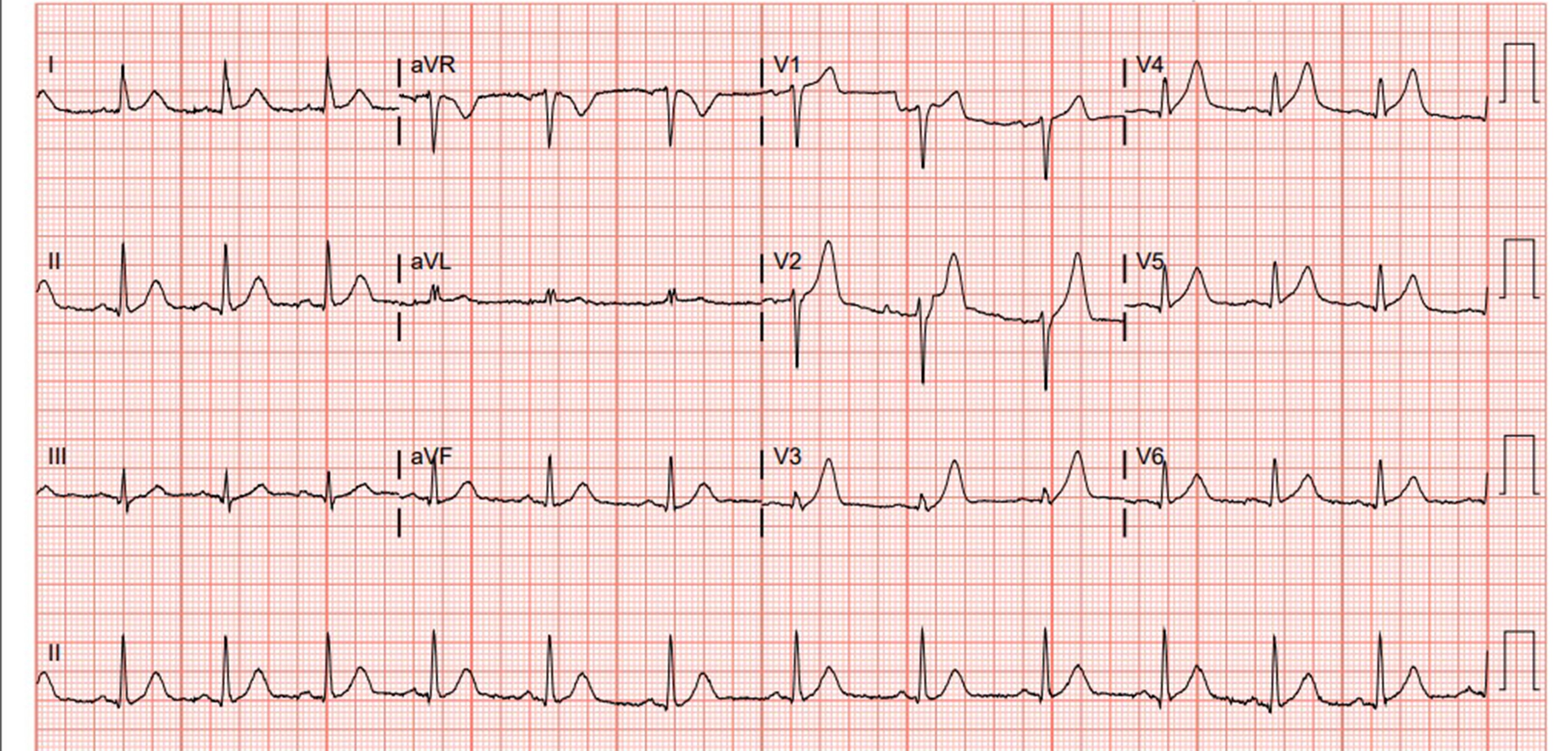
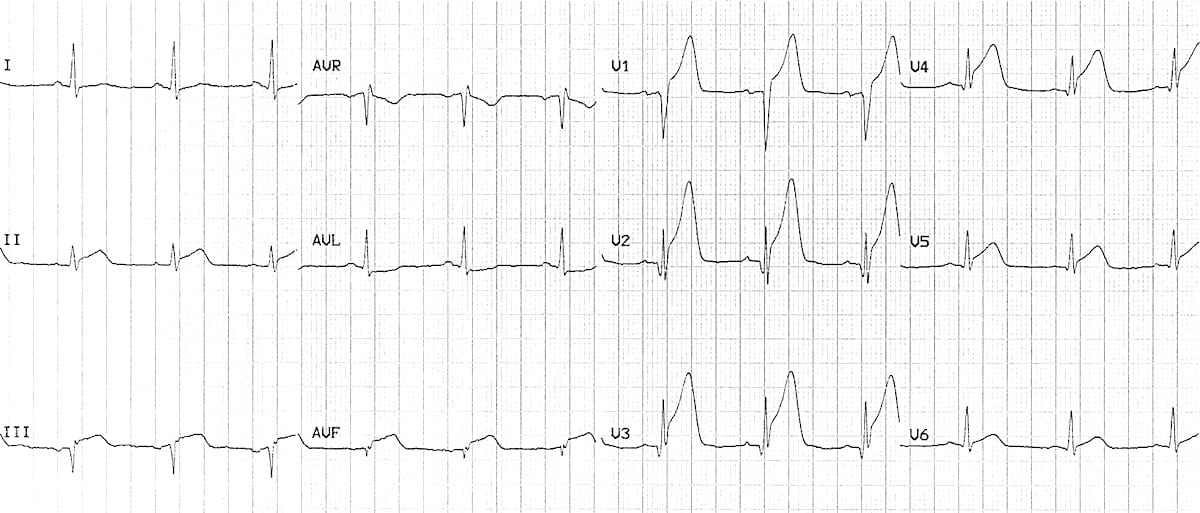
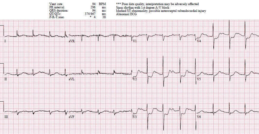
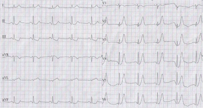
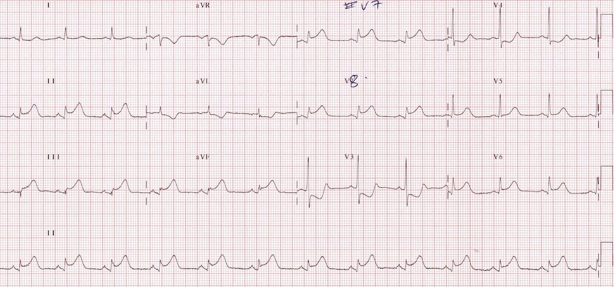
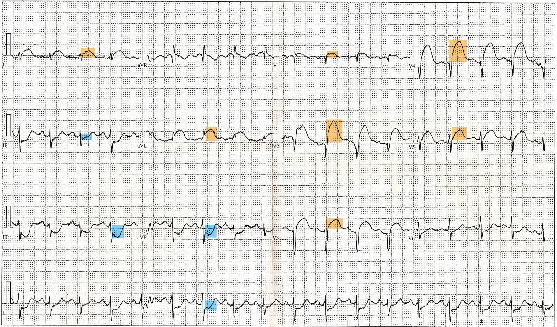
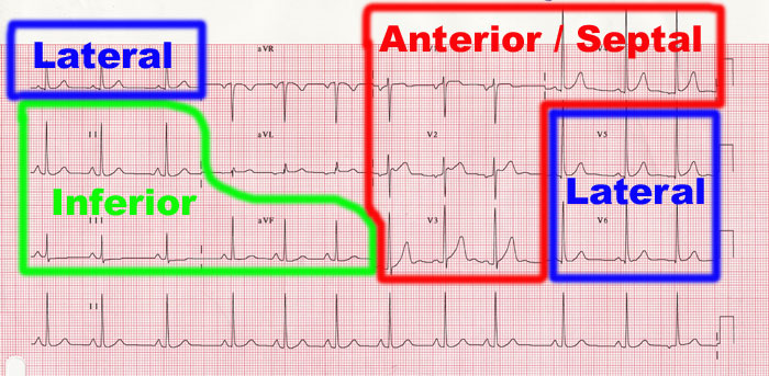
Diagnosing an acute myocardial infarction by ecg is an important skill for healthcare professionals mostly because of the stakes involved for the patient.
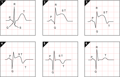
Mi on ecg. Explanation of the ecg changes in v1 3. Therefore it may be difficult to estimate the duration of the ischemia on the ecg which is crucial for adequate treatment. St elevation becomes st depression. Symptoms patients with acute myocardial infarction may present with typical ischemic chest pain or with dyspnea nausea unexplained weakness or a combination of these symptoms.
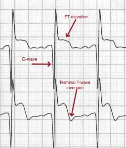
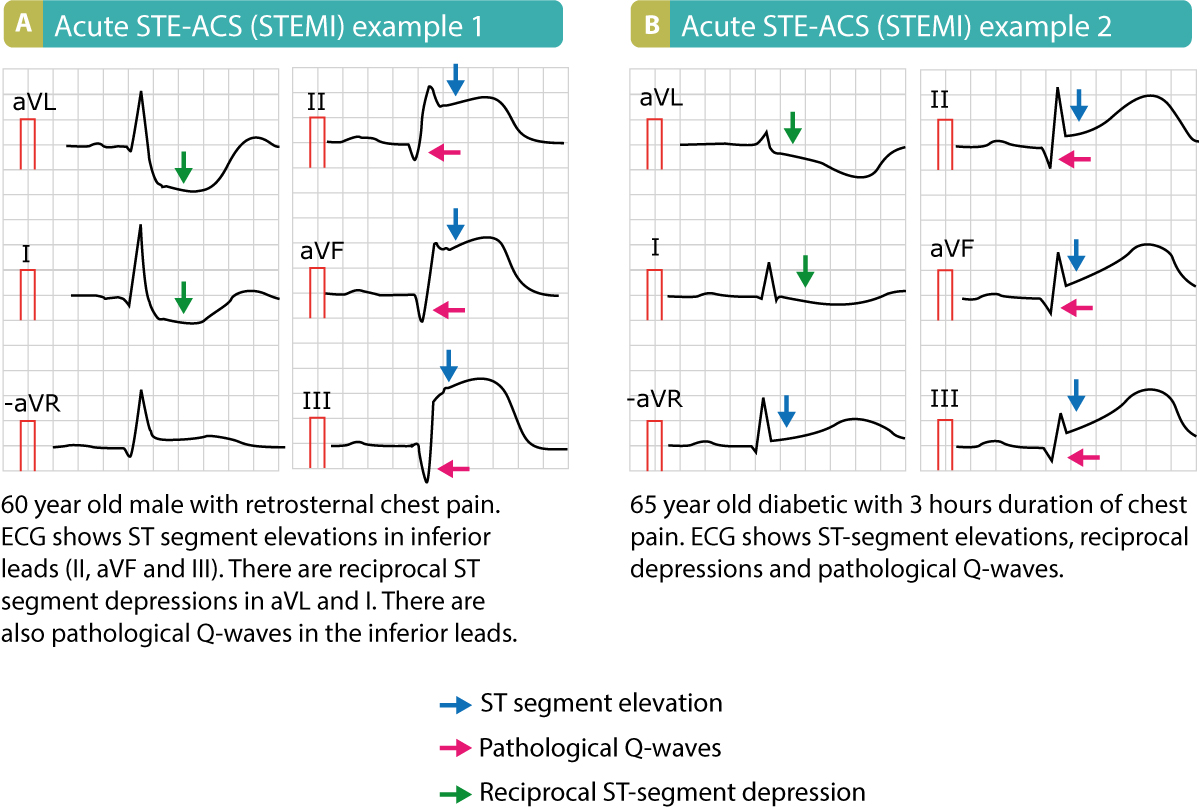
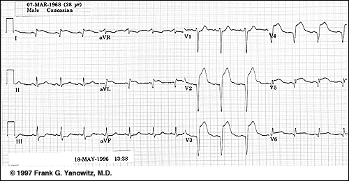
2015 acc aha scai focused update on primary pci for patients with stemi. Mi s resulting from total coronary occlusion result in more homogeneous tissue damage and are usually reflected by a q wave mi pattern on the ecg. One of the complications with using ecg for myocardial infarction diagnosis is that it is sometimes difficult to determine which changes are new and which are old. Ecg is the mainstay of diagnosing stemi which is a true medical emergency making the correct diagnosis promptly is life saving if the clinical picture is consistent with mi and the ecg is not diagnostic serial ecg at 5 10 min intervals several conditions can be associated with st elevation on ecg most commonly lbbb pericarditis and early repolarization if in doubt call the cardiologist or activate the cath lab.
Mi s resulting from subtotal occlusion result in more heterogeneous damage which may be evidenced by a non q wave mi pattern on the ecg. Signs and symptoms of myocardial ischemia. The 12 lead ecg is used to classify mi patients into one of three groups. Missing a st segment.
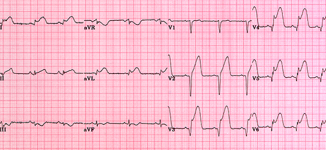
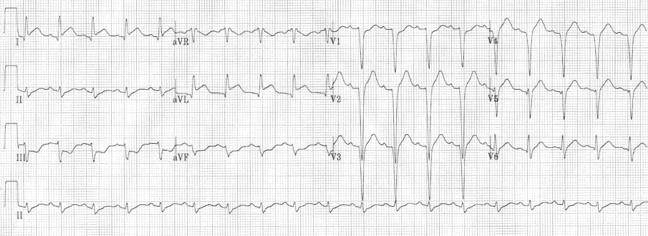
Those with st segment elevation or new bundle branch block suspicious for acute injury and a possible candidate for acute reperfusion therapy with thrombolytics or primary pci those with st segment depression or t wave inversion suspicious for ischemia and. Because posterior electrical activity is recorded from the anterior side of the heart the typical injury pattern of st elevation and q waves becomes inverted. While the ischemia lasts several ecg changes will occur and disappear again. The anteroseptal leads are directed from the anterior precordium towards the internal surface of the posterior myocardium.
Identifying an acute myocardial infarction on the 12 lead ecg is the most important thing you can learn in ecg interpretation. Severe ischemia results in ecg changes within minutes.


